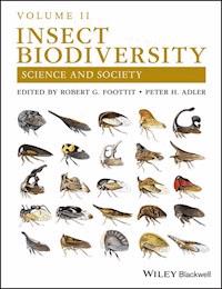
459,99 zł
Volume Two of the new guide to the study of biodiversity in insects Volume Two of Insect Biodiversity: Science and Society presents an entirely new, companion volume of a comprehensive resource for the most current research on the influence insects have on humankind and on our endangered environment. With contributions from leading researchers and scholars on the topic, the text explores relevant topics including biodiversity in different habitats and regions, taxonomic groups, and perspectives. Volume Two offers coverage of Insect Biodiversity in regional settings, such as the Arctic and Asia, and in particular habitats including crops, caves, and islands. The authors also include information on historical, cultural, technical, and climatic perspectives of Insect Biodiversity. This book explores the wide variety of insect species and their evolutionary relationships. Case studies offer assessments on how Insect Biodiversity can help meet the needs of a rapidly expanding human population, and examine the consequences that an increased loss of insect species will have on the world. This important text: * Offers the most up-to-date information on the important topic of Insect Biodiversity * Explores vital topics such as the impact on Insect Biodiversity through habitat loss and degradation and climate change * With its companion Volume I, presents current information on the biodiversity of all insect orders * Contains reviews of Insect Biodiversity in culture and art, in the fossil record, and in agricultural systems * Includes scientific approaches and methods for the study of Insect Biodiversity The book offers scientists, academics, professionals, and students a guide for a better understanding of the biology and ecology of insects, highlighting the need to sustainably manage ecosystems in an ever-changing global environment.
Ebooka przeczytasz w aplikacjach Legimi na:
Liczba stron: 2413
Cover
List of Contributors
Foreword
Preface, Volume II
Acknowledgments
Chapter 1: Introduction – A Brief History of Revolutions in the Study of Insect Biodiversity
1.1 Discovery
1.2 Conceptual Development
1.3 Information Management
1.4 Conclusions
Acknowledgments
References
Part I Habitats and Regions
Chapter 2: Insect Biodiversity in the Arctic
2.1 Documenting Biodiversity – Traditional Taxonomy Versus DNA Barcoding
2.2 Insect Species Diversity in the Arctic
2.3 Historical Insect Biodiversity in the Arctic – the Time Perspective
2.4 Biodiversity on the Landscape Scale
2.5 Important Characteristics of Arctic Insect Biodiversity
2.6 Cold Tolerance – a Diversity of Adaptations
2.7 Dispersal, Immigration, and Biodiversity
2.8 Pollinator Networks and Pollinator Biodiversity
2.9 A Biodiversity Paradise for Parasites?
2.10 Biodiversity and the Changing Arctic Climate
References
Chapter 3: Insect Biodiversity in Indochina: A Window into the Riches of the Oriental Region
3.1 Physical Geography and Climate
3.2 Features of Insect Biodiversity in the Lower Mekong Subregion
3.3 Insect Biodiversity and Society in Indochina
3.4 Conclusions
Acknowledgments
References
Chapter 4: Biodiversity of Arthropods on Islands
4.1 What is an Island?
4.2 Ecological Attributes of Islands
4.3 Evolution on Islands
4.4 Evolution in Other Insular Environments
4.5 Characteristics of Island Biodiversity
4.6 Conservation
4.7 Conclusion
References
Chapter 5: Beneficial Insects in Agriculture: Enhancement of Biodiversity and Ecosystem Services
5.1 Components of Biodiversity: Species Richness, Species Evenness, and Species Identity
5.2 Why Does Insect Biodiversity Matter to Agriculture?
5.3 Degradation of Biodiversity Through Agricultural Intensification, and Its Reversal
5.4 Restoring Biodiversity to Agroecosystems
5.5 Conclusions and Recommendations
5.6 Summary
Acknowledgments
References
Chapter 6: Insects in Caves
6.1 The Story of
Leptodirus hochenwartii
6.2 The Variety of Subterranean Spaces
6.3 Ecological Roles of Insects in Caves
6.4 Morphological and Life‐History Adaptations of Insects to Subterranean Life
6.5 Probable Modes of Successful Colonization of Subterranean Space
6.6 Taxonomic and Geographic Patterns of Subterranean Insect Biodiversity
6.7 Human Utility and Protection of Cave Insects
References
Part II: Taxa
Chapter 7: Biodiversity of the Thysanurans (Microcoryphia and Zygentoma)
7.1 Paleontological Data
7.2 Parasitism
7.3 Predation
7.4 Order Microcoryphia (= Archaeognatha)
7.5 Order Zygentoma (= Thysanura
Sensu Stricto
)
7.6 Genetic Studies of Thysanurans
7.7 Thysanurans and Humans
7.8 Geographic Distribution of the Thysanurans
References
Chapter 8: Biodiversity of Zoraptera and Their Little‐Known Biology
8.1 Morphology
8.2 Life History and Ecology
8.3 Reproduction
8.4 Phylogenetic Position – “The Zoraptera Problem”
8.5 Conclusion
Acknowledgments
References
Chapter 9: Biodiversity of Embiodea
9.1 Diversity in Habitat and Silk
9.2 The Promise of Silk‐Like Biomaterials and Emerging Lessons from Webspinners
9.3 Social Behavior
9.4 Families of Embiodea
9.5 Webspinners of the Fossil Record
9.6 Conclusion
References
Chapter 10: Biodiversity of Orthoptera
10.1 Taxonomic Classification and Phylogeny
10.2 Diversity and Distribution
10.3 Morphological and Biological Diversity
10.4 Societal Importance
10.5 Overview of Taxa
Acknowledgments
References
Chapter 11: Biodiversity of Phasmatodea
11.1 Phasmatodean Phylogeny
11.2 Overview of Taxa
11.3 The Phasmatodean Fossil Record
11.4 Phasmatodea as Research Tools
11.5 Importance to Human Society
References
Chapter 12: Biodiversity of Dermaptera
12.1 Epizoic Dermaptera
12.2 Structure and Function
12.3 Locomotion
12.4 Distribution
12.5 Development and Reproduction
12.6 Behavior
12.7 Parasitism and Symbiosis
12.8 Fossils and Research History
12.9 Overview of Taxa
12.10 Societal and Scientific Importance
Acknowledgments
References
Chapter 13: Biodiversity of Grylloblattodea and Mantophasmatodea
Grylloblattodea
13.2 Mantophasmatodea
13.3 Fossil Record
13.4 Conclusions
Acknowledgments
References
Chapter 14: Biodiversity of Blattodea – the Cockroaches and Termites
14.1 Overview of Taxa
14.2 Societal Importance
References
Chapter 15: Biodiversity of Mantodea
15.1 Morphological and Biological Diversity
15.2 Phylogeny and Classification
15.3 Morphological Convergence and Ecomorphs
15.4 Conclusions
References
Chapter 16: Biodiversity of Psocoptera
16.1 Classification
16.2 Overview of the Psocoptera
16.3 Summary of Diversity of the Psocoptera and Predictions
16.4 The Importance to Humans of Psocopteran Biodiversity
Acknowledgments
References
Chapter 17: Biodiversity of Ectoparasites: Lice (Phthiraptera) and Fleas (Siphonaptera)
17.1 Phthiraptera – TheParasitic Lice
17.2 Siphonaptera – The Fleas
17.3 Medical and Veterinary Importance
17.4 Community Diversity of Lice and Fleas
17.5 Conservation of Lice and Fleas
Acknowledgments
References
Chapter 18: Biodiversity of Thysanoptera
18.1 What Are Thrips?
18.2 Family Diversity
18.3 The Lives of Thrips
18.4 Thrips Around the World
18.5 Thrips as Research Targets
18.6 Structural Diversity of Thrips
18.7 Thrips as Pests
18.8 Thrips and Human Life
18.9 Thrips Information Sources
References
Chapter 19: The Diversity of the True Hoppers (Hemiptera: Auchenorrhyncha)
19.1 Overview of the Auchenorrhyncha
19.2 Prospectus
Acknowledgments
References
Addendum
Chapter 20: The Biodiversity of Sternorrhyncha
20.1 Sternorrhyncha and Society
20.2 Taxonomic Diversity of Sternorrhyncha
20.3 Functional Diversity of Sternorrhyncha
20.4 Conclusions
Acknowledgments
References
Chapter 21: Biodiversity of the Neuropterida (Insecta: Neuroptera, Megaloptera, and Raphidioptera)
21.1 Phylogeny
21.2 Geological Age
21.3 Metamorphosis and Life Stages
21.4 Biology
21.5 Distribution
21.6 Overview of Orders and Families
21.7 Societal Importance
21.8 Scientific Importance
Acknowledgments
References
Chapter 22: Biodiversity of Strepsiptera
22.1 Family Bahiaxenidae
22.2 Suborder Mengenillidia
22.3 Suborder Stylopidia
22.4 Infraorder Stylopiformia
22.5 Conclusions
Acknowledgments
References
Chapter 23: Biodiversity of Mecoptera
23.1 Suborder Nannomecoptera
23.2 Suborder Pistillifera
23.3 Societal Value of Mecoptera
23.4 Scientific Value of Mecoptera
23.5 Conclusion
References
Part III: Perspectives
Chapter 24: The Fossil History of Insect Diversity
24.1 Importance of the Insect Fossil Record
24.2 Types of Insect Diversity Past and Present
24.3 Biodiversity Changes Through Time
24.4 Current Societal Aspects of Fossil Insect Biodiversity
24.5 Conclusions
Acknowledgments
References
Chapter 25: Phenotypes in Insect Biodiversity Research
25.1 Phenotype Data: Past and Present
25.2 Phenotype Data: Present and Future
25.3 Challenges and Future Directions
Acknowledgments
References
Chapter 26: Global Change and Insect Biodiversity in Agroecosystems
26.1 Global Change
26.2 Insect Biodiversity in Agriculture
26.3 Effects of Global Change on Biodiversity – What Do We Know?
26.4 Island Versus Continent Contrasts
26.5 Tropical Versus Temperate Issues
26.6 Some Concluding Viewpoints
References
Chapter 27: Digital Photography and the Democratization of Biodiversity Information
27.1 The Digital Insect Collection
27.2 Digital Images in Interactive Keys
27.3 Digital Photography and Taxonomic Revisions
27.4 Organization of Digital Insect Collections
27.5 Conclusions
References
Chapter 28: Bee (Hymenoptera: Apoidea: Anthophila) Diversity Through Time
28.1 Morphological Diversity
28.2 Behavioral Diversity: Social, Nesting, and Floral Hosts
28.3 Geographical Diversity
28.4 Evolutionary History and Diversification
28.5 Conclusions
References
Chapter 29: Insect Biodiversity in Culture and Art
29.1 Prehistory
29.2 Insects in the Ancient World
29.3 The Cult of Artemis: A Case Study
29.4 Roman Insect Art
29.5 Ancient China
29.6 Religions of India
29.7 Post‐Classical Era
29.8 The Americas
29.9 Modern History
29.10 Japanese Art
29.11 Language and Literature
29.12 Insects in Music
29.13 Insects in Cinema
29.14 Akihabara Culture: Toys, Video Games, and Anime from Modern Japan
29.15 Present and Future Trends in Cultural Entomology
29.16 The Internet Age
References
Index of Arthropod Taxa Arranged by Order and Family
Index of Arthropod Taxa Arranged Alphabetically
Index of non‐Arthropod Taxa Arranged Alphabetically
Subject Index
End User License Agreement
Chapter 02
Table 2.1 Comparison of insect species diversity expressed as the number of genera and species of insects in each family in the High and Low Arctic Regions of North America.
Table 2.2 Recorded insect biodiversity of the High‐Arctic Islands of the Barents Sea.
Table 2.3 Recorded insect species diversity of aquatic and humid habitats in subarctic Finnmark, Scandinavia.
Table 2.4 Vegetation types near Ny‐Ålesund, Svalbard, with their dominant terrestrial Chironomidae species.
Table 2.5 Diptera families and species involved in the decomposition of organic matter on Svalbard.
Table 2.6 Habitat preferences of Hemiptera species found on Dolgii Island (Barents Sea) in various parts of their broader distribution.
Chapter 03
Table 3.1 Numbers of insect species in each order for countries of the Lower Mekong Subregion.
Table 3.2 Number of insect species and genera, excluding Coleoptera, in the Lower Mekong Subregion.
Chapter 06
Table 6.1 Number of specialized cave insects, soil insects, and generalists at a milieu souterrain superficiel (MSS) site in Teno, Tenerife, Canary Islands.
Table 6.2 Summary of environmental variability of shallow subterranean habitats, based on hourly records, mostly of temperature, and comparison to values at surface.
Table 6.3 Comparison of classification terminology for ecological categories.
Table 6.4 Ratios of antennal length, tarsus‐three length, and overall length index to pronotal length for Leiodidae beetles in the genus
Ptomaphagus
, with the mean value for the epigean species subtracted.
Table 6.5 Life‐history characteristics of various
Ptomaphagus
(Leiodidae) beetle species reared in the laboratory.
Table 6.6 Parapatric cave and surface populations on Hawaii Island.
Table 6.7 Caves with more than 25 troglobionts.
Chapter 07
Table 7.1 Recent families, subfamilies, and genera
incertae sedis
in the Microcoryphia (after Sturm and Bach de Roca 1993 and Kaplin 2000).
Table 7.2 Recent genera of Microcoryphia (including paleoforms), number of subgenera (Subg.) and recent species (Spp.), and their distributions.
Table 7.3 Families and subfamilies in the Zygentoma.
Table 7.4 Recent genera of families in the order Zygentoma (fossil family Lepidotrichidae excluded), number of described species (Spp.), and their distribution by zoogeographical region and continent.
Chapter 08
Table 8.1 Checklist of described Zoraptera.
Chapter 09
Table 9.1 Number of extant genera and species in families of the order Embiodea.
Chapter 10
Table 10.1 Summary of the known number of orthopteran subfamilies, genera, and species for each family.
Chapter 11
Table 11.1 Biodiversity of major phasmatodean groups, largely following the arrangement of Brock et al. (2016), but modified according to several phylogenetic results not reflected in this databank and omitting subspecies.
Chapter 12
Table 12.1 Families and described, valid species of living Dermaptera.
Chapter 13
Table 13.1 Extant species and subspecies of Grylloblattodea.
Table 13.2 Extant and fossil species of Mantophasmatodea.
Chapter 14
Table 14.1 Major groups of Blattodea (cockroaches including termites) with numbers of known species.
Table 14.2 Cockroach classification.
Chapter 15
Table 15.1 Current classification of the Mantodea, with genus and species numbers of all currently recognized subgroups.
Table 15.2 Current classification of the Mantodea, with an overview of genus and species numbers in each family.
Chapter 16
Table 16.1 Number of genera and species of Psocoptera by family for major biogeographic divisions of the world (Divisions I, II, VI, VII, VIII, and IX primarily Eastern Hemisphere).
Table 16.2 Number of genera and species of Psocoptera by family for major biogeographic divisions of the world (Divisions III, IV, V, and X, primarily Western Hemisphere, and the world).
Chapter 17
Table 17.1 Suborders and families of lice (Phthiraptera) with approximate numbers of known genera and species in each family, as of 2014.
Table 17.2 Families and subfamilies of fleas (Siphonaptera) with approximate numbers of known genera and species in each, as of 2014.
Chapter 18
Table 18.1 Classification of the Thysanoptera as of January 2018.
Chapter 19
Table 19.1 Distribution of Auchenorrhyncha genera and species by higher taxon (to subfamily) and zoogeographic region.
Table 19.2 Summary of available data on life‐history patterns in the Membracidae (Wood 1984; Lin et al. 2004; Wallace and Deitz 2004; Albertson and Dietrich 2005; Costa 2006; Godoy et al. 2006; Wallace 2011; O. Evangelista, personal communication on Heteronotinae).
Table 19.3 Summary of Fulgoromorpha biodiversity and species density by zoogeographic region.
Table 19.4 Summary of Cicadomorpha biodiversity and species density by zoogeographic region.
Chapter 20
Table 20.1 Taxonomic diversity of sternorrhynchan endosymbionts.
Chapter 21
Table 21.1 Primary natural activity period and relative frequency of nocturnal light attraction by adults of each family of the Neuropterida.
Table 21.2 An alphabetical list of the orders and families of the extant Neuropterida of the world (after Oswald 2015), with counts of genera and species.
Table 21.3 A higher classification (order to tribe) of the extant Neuropterida of the world (after Oswald 2015).
Chapter 22
Table 22.1 Hierarchical classification of Strepsiptera.
Table 22.2 Comparison of koinobionts, idiobionts (Askew and Shaw 1986, Strand 1986, Gauld and Bolton 1988, Godfray 1994, Strand and Peach 1995, Pennacchio and Strand 2006), and Strepsiptera (not in order of importance) (after Kathirithamby 2009).
Table 22.3 Families and genera of Strepsiptera, with species numbers and hosts.
Chapter 23
Table 23.1 Number of extant genera and species of Mecoptera by family.
Chapter 24
Table 24.1 Biological and geological scale of 30 biodiversity studies using the plant–insect fossil record from the Labandeira, Wilf and Wappler Laboratories
*
.
Chapter 25
Table 25.1 Some ontologies relevant to insect biodiversity research; note that several other ontologies are available or required for proper formalization of insect phenotype data.
Table 25.2 Typical character statements and their semantic representations.
Chapter 28
Table 28.1 Numbers of species and mean and 95% highest posterior density interval (HPD) of crown ages and stem ages of bee lineages.
Chapter 29
Table 29.1 The earliest recorded English use of insect nouns as recorded in the
Oxford English Dictionary Online
(http://www.oed.com/). The year of first use precedes the English word.
Chapter 01
Figure 1.1 Selected highlights in the insect biodiversity time line.
Chapter 02
Figure 2.1 Map showing the major regions of the Arctic and some of the important sites named in the text. Image created by author.
Figure 2.2 The changing relative percentages of herbivorous, aquatic, and other terrestrial insect species groups, with respect to increasing climate severity in the Arctic regions of North America (based on Danks 1992b; redrawn from Hodkinson et al. 2013). At high latitudes, aquatic species are predominantly Diptera with aquatic stages and water beetles.
Chapter 03
Figure 3.1 Map of Indochina showing the location of the Mekong River. Dashed line and arrows indicate the separation point of the upper and lower Mekong. Author originated. Base map exported from R package
maps
(Original S code by Richard A. Becker, Allan R. Wilks. R version by Ray Brownrigg. Enhancements by Thomas P Minka and Alex Deckmyn. (2016). maps: R package version 3.1.0.)
Figure 3.2 Diversity of insect species from the Lower Mekong Subregion. (a)
Papilio
sp. (Lepidoptera). (b)
Graphium antiphates
(Lepidoptera). (c)
Apis dorsata
(Hymenoptera). (d)
Xylocopa
sp. (Hymenoptera). (e)
Cucujus
sp. (Coleoptera). (f)
Aphis nerii
(Hemiptera). Images by authors.
Figure 3.3 Insect foods in local market of Laos. (a) Coleoptera. (b) bamboo caterpillar (Lepidoptera). (c−g) Orthoptera. (h) Hemiptera. Images by authors.
Chapter 04
Figure 4.1 Different kinds of insular systems. (a) Oceanic island. The island of Pico in the archipelago of the Azores. This archipelago is one of the best studied for arthropods, as evidenced through the work of Borges and colleagues (Borges 1992, Borges and Brown 1999, Cardoso et al. 2010, Triantis et al. 2010a, Triantis et al. 2010b, Cardoso et al. 2011, Gaspar et al. 2011, Meijer et al. 2011). (b) Cave. View towards entrance from within Algar do Carvão, an ancient lava tube on the Azorean island of Terceira, which harbors a number of endemics, including both spiders and insects (Reboleira et al. 2011). (c) Pleistocene fragment island. Agistri in the Sarconic Islands of Greece. Early on (23–12 million years ago), the Greek islands were all connected in a continuous land mass. Sea transgression (12–5 mya) formed a mid‐Aegean barrier, followed by fragmentation and widening of the Aegean, leading to the Pleistocene, which was characterized by eustatic sea‐level change (Triantis and Mylonas 2009). The long history of connection and isolation has shaped the diversity of arthropods known from the region today (Sfenthourakis and Legakis 2001). (d) Forest fragment. Shown is a “kipuka,” or island of forest surrounded by lava, on the island of Hawaii. Arthropods are often isolated in these fragments and show clear genetic differences among kipukas (Vandergast and Gillespie 2004, Vandergast et al. 2004). (e) Unique habitat islands. Mono Lake is an example of a unique habitat – a saline lake – that is isolated from similar such habitats. It harbors distinct assemblages of organisms, particularly notable being the brine shrimps and alkali flies (Herbst 1999). Other habitat types that hold unique assemblages of arthropods include sand dunes (Van Dam and Matzke 2016) and vernal or desert pools (Ward and Blaustein 1994). (f) Recent fragment islands. Barro Colorado island, in Gatun Lake of Panama, was formed by flooding of the Chagres River in the creation of the Panama Canal (Leigh 2009). As a result, species numbers declined through relaxation of the supersaturated insular biota as it returns to equilibrium. A similar phenomenon has been documented for oaks, which act as islands for leaf‐mining insects (Opler 1974). (g) Sky islands. The American Madrean sky islands of southeastern Arizona and New Mexico have served to isolate many arthropods on the mountain summits. Particularly well known are the jumping spiders in the
Habronattus pugillis
complex (Masta 2000), scorpions (Hughes 2011), and beetles (Smith and Farrell 2005, Ober and Connolly 2015). All photographs by George K. Roderick, used with permission. (
See color plate section for the color representation of this figure
.)
Chapter 05
Figure 5.1 Species richness describes the number of different species present, irrespective of abundance, with low species richness (a) characterized by few species, and high species richness (b) characterized by many species. Species evenness describes the relative abundances of species in an assemblage, with low evenness (c) characterized by relatively more of particular species than others, and high evenness (d) characterized by relatively even abundances of the species present. Here we illustrate how, even when the number of individuals in an assemblage is held constant, levels of species richness and evenness can vary dramatically. Original figure by author.
Figure 5.2 Temporal and spatial complementarity. (a–c) Temporal complementarity is the mechanism by which a lepidopteran corn pest (
Helicoverpa zea
) is most effectively controlled (Pfannenstiel and Yeargan 2002). During the day, a lady beetle is an effective predator of the moth eggs (a); at night, a predatory bug is an effective egg predator (b). Predation is maximized through the combined effects of two natural enemies; only with both enemies present are the moths deprived of a daily (temporal) refuge from predation (c). (d–f) On collard plants, spatial complementarity among predator species leads to effective control of pest aphids only when multiple predator species co‐occur (Straub and Snyder 2008). This level of control is because some predator species forage mostly on leaf edges (d), while other predator species access aphids at the center of leaves (e). Only with a diverse predator community (f) are all of the spatial refuges of the aphids removed. Original figure by author.
Figure 5.3 Two extremes of land‐use intensification. (a) Monoculture landscape growing leafy greens in the Central Valley in California, USA. This type of large‐scale farming relies heavily on synthetic fertilizers, tillage, and chemical control of pest insects. There is an obvious dearth of resources that might benefit insects. (b) Highly diversified farm with multiple crops, orchards, livestock, and weedy field edges, all of which can contribute to a healthier, more diversified insect community. Original figure by author.
Chapter 06
Figure 6.1 Photograph of
Leptodirus hochenwartii
, the first insect described from caves. Photo by Slavko Polak, used with permission.
Figure 6.2 Range map of
Leptodirus hochenwartii
in Slovenia, courtesy of Slavko Polak. The range of
L. hochenwartii
in neighboring Croatia is approximately the same size, with three additional subspecies present.
Figure 6.3 Histogram of log of pore (habitat) size for different shallow subterranean habitats. MSS, milieu souterrain superficiel. From Culver and Pipan (2014), by permission of Oxford University Press.
Figure 6.4 Comparative normalized spatial density map of 19 cave‐limited arthropods (mostly insects) with respect to vertical distance from the surface in small caves in northeastern Slovenia. Adapted from Novak et al. (2012).
Figure 6.5 Photo of exposed milieu souterrain superficiel (MSS) in a
Fagus
forest in Mašun, Slovenia. Photo by T. Pipan.
Figure 6.6 Conceptual diagram of different terrestrial shallow subterranean habitats. The position of the rectangle indicates the typical range of pore (habitat) size and proximity to the soil. MSS, milieu souterrain superficiel. From Culver and Pipan (2014), by permission of Oxford University Press.
Figure 6.7 Parallel evolution in caves of the foot complex of cave Collembola. (a)
Pseudosinella
in the United States. (b–d) Different lineages of
Pseudosinella
in Europe. (e,f) Different lineages of
Sinella
in the United States. (g)
Sinella
in Japan. The least troglomorphic species are shown at the top and the most troglomorphic species below. From Christiansen (2012), by permission of Elsevier.
Figure 6.8 Latitudinal variation in divergence times of eight sympatric sister pairs of stygobiotic dytiscid beetles in Western Australia. The open circles show species pairs belonging to the Bidessini, the black circles show species pairs belonging to the Hydroporini. From Leys et al. (2003).
Figure 6.9 Pie diagrams of relative contributions of insects and other major groups to species richness of three of the seven hotspot caves in Table 6.7 (Cueva de Filipe Reventón, Canary Islands; Mammoth Cave, Kentucky, United States; and Vjetrenica, Bosnia and Herzegovina). Data from various sources. Original figure by authors.
Figure 6.10 (a) Map of species richness patterns of US troglobionts. Major karst areas of the eastern and central United States are shown in light gray. Although there are many caves in the western United States and Canada, there are no large karst areas, and no areas of rich fauna. The open triangles are areas with few if any troglobionts, and the gray triangles are areas with fewer than 50 species, usually many fewer than 50. The three open circles are areas analyzed in this study with less than 50 species in an area of 5000 km
2
or less. The black circle is the diversity hotspot in northeastern Alabama. The boundary of the Pleistocene ice sheet is shown as a solid line. A pair of dashed lines indicates the hypothesized position of the high‐diversity ridge. (b) Map of species‐richness patterns of European troglobionts. The open triangles are areas with few if any troglobionts, the gray triangles are areas with fewer than 50 species, usually many fewer than 50, and the gray circle is Ardèche, with fewer than 50 species in an area of 5000 km
2
or less. The black circles are the diversity hotspots in Slovenia and Ariège. Black triangles are other possible diversity hotspots. The boundary of the Pleistocene ice sheet is shown as a scored solid line. A pair of dashed lines indicates the hypothesized position of the high‐diversity ridge. Adapted from Culver et al. (2006).
Figure 6.11 Proportion of troglobiotic species in different families of cave beetles. From Moldovan (2012), by permission of Elsevier.
Chapter 07
Figure 7.1 Dominican amber fossils (20–25 million years old) of Zygentoma. (a)
Archeatelura sturmi
Mendes, 1997b (Zygentoma: Nicoletiidae). (b)
Ctenolepisma electrans
Mendes, 1998a (Zygentoma: Lepismatidae). Photographs by author.
Figure 7.2 Microcoryphia. (a) Body of Microcoryphia in dorsal view. After Handschin (1929). (b) Body of Microcoryphia in lateral view. After Handschin (1929). (c) Head of
Haslundichilis
(Microcoryphia: Machilidae), frontal view. After Mendes (1982). (d) Head of
Pedetontus
(Microcoryphia: Machilidae), frontal view. After Mendes (1992). (e) Head of
Bachilis
(Microcoryphia: Machilidae), frontal view. After Mendes (1977). (f) Monocondylian mandible of Microcoryphia. Original. (g) Maxillary palp of male of
Promesomachilis
(Microcoryphia: Machilidae). Original. (h) Maxillary palp of male of
Machilinus
(Microcoryphia: Meinertellidae). After Mendes (2005). (i). Labial palp of male of
Promesomachilis
(Microcoryphia: Machilidae). Original. (j). Median and hind legs of
Dilta
(Microcoryphia: Machilidae), both with coxal stylets. After Mendes et al. (1996). (k) Median and hind legs of
Machilontus
(Microcoryphia: Meinertellidae); coxal stylets only on hind legs. Original. (l) Median (left) and hind (right) legs of
Machilinus
(Microcoryphia: Meinertellidae); both are devoid of coxal stylets. Original.
Figure 7.3 Zygentoma. (a) Coxa and coxal stylet of male hind leg of
Mesomachilis nearcticus
(Microcoryphia:
incertae sedis
). After Mendes (1992). (b) Coxa and coxal stylet of female hind leg of
Mesomachilis nearcticus
(Microcoryphia:
incertae sedis
). Original. (c) Median abdominal coxosternite (urosternite) of
Machilinus
(Microcoryphia: Meinertellidae), with the sternite extremely reduced. After Mendes (2005). (d) Median abdominal coxosternite (urosternite) of
Dilta
(Microcoryphia: Machilidae), with only one pair of coxal vesicles (arrow) beyond the abdominal stylets. Original. (e) Median urosternite (coxosternite) of
Machilis
(Microcoryphia: Machilidae), with two pairs of coxal vesicles (arrows) beyond the abdominal stylets. Original. (f) Coxites VIII (left) and IX (right) of male of
Haslundichilis
(Microcoryphia: Machilidae) and genitalia (penis and two pairs of paramera). After Mendes (1982). (g) Coxites VIII (left) and IX (right) of male of
Allopsontus
(
Machilanus
) (Microcoryphia: Machilidae) and genitalia (penis plus paramera IX only). Original. (h) Coxite IX of male of
Machiloides
(Microcoryphia: Meinertellidae) and genitalia (penis only, paramera lacking). After Mendes (1992). (i) Ovipositor of the primary type. Apex of gonapophyses VIII (left) and IX (right) of female of
Promesomachilis
(Microcoryphia: Machilidae). Original. (j) Ovipositor of the secondary type. Apex of gonapophyses VIII (left) and IX (right) of female of
Coreamachilis
(Microcoryphia: Machilidae). After Mendes (1993). (k) Ovipositor of the tertiary type; median and terminal divisions of gonapophyses VIII (left) and IX (right) of female of
Machilontus
(Microcoryphia Meinertellidae). After Mendes (1989c). (l) Ovipositor of the quaternary type; median and terminal divisions of gonapophyses VIII (left) and IX (right) of female of
Machilinus
(Microcoryphia, Meinertellidae). After Mendes (2005).
Figure 7.4 Zygentoma. (a) Profile of the common house silverfish,
Lepisma saccharina
(Zygentoma: Lepismatidae), dorsal view. After Handschin (1929). (b) Profile of the common house silverfish
L. saccharina
(Zygentoma: Lepismatidae), lateral view. After Handschin (1929). (c) Profile of
Allonicoletia
(Zygentoma: Nicoletiidae: Cubacubaninae). After Mendes (1992). (d) Profile of a female of the termitophilous
Allomorphuroides
(Zygentoma: Nicoletiidae: Atelurinae). After Mendes (1987a). (e) Head (right half) of
Thermobia
(Zygentoma: Lepismatidae). Original. (f) Head of
Allonicoletia
(Zygentoma: Nicoletiidae). After Mendes (1992). (g) Dicondylian mandible of Zygentoma. Original. (h) Maxillary palp of
Neoasterolepisma
(Zygentoma: Lepismatidae). Original. (i) Labial palp of
Neoasterolepisma
(Zygentoma: Lepismatidae). Original. (j) Apex of maxillary palp (distal division) of
Neoasterolepisma
(Zygentoma: Lepismatidae). After Mendes (1991). (k) Apex of maxilla of
Allonicoletia
(Zygentoma: Nicoletiidae) with prostheca. After Mendes (1992). (l) Apex of maxilla of
Protrinemuroides
(Zygentoma: Protrinemuridae) devoid of prostheca. Original. (m) Leg of
Ctenolepisma
(Zygentoma: Lepismatidae). After Mendes (1980b).
Figure 7.5 Typical pretarsus of a silverfish, with two claws and a claw‐like empodium of
Neoasterolepisma
(Zygentoma: Lepismatidae). Scanning electron micrograph by author.
Figure 7.6 Zygentoma. (a) Entire median coxosternite of
Neoasterolepisma
(Zygentoma: Lepismatidae). After Mendes (1980b). (b) Median coxosternite of
Anelpistina
(Zygentoma: Nicoletiidae), divided into a median sternite plus 1 + 1 lateral coxites. Original. (c) Elongated and annulated paramera of male of
Allacrotelsa
(Zygentoma: Lepismatidae). Original. (d) Thin and tubular paramera (entire) of male of
Acrotelsa
(Zygentoma: Lepismatidae). Original. (e) Vesiculiform medium‐size paramera of male of
Neoasterolepisma
(Zygentoma: Lepismatidae). After Mendes (1980b). (f) Cylindrical paramera IX of
Santhomesiella
(Zygentoma: Nicoletiidae). After Mendes (1988a). (g) Vesiculiform large paramera of male of
Tricholepisma
(Zygentoma: Lepismatidae). Original. (h) Absence of paramera in male of
Ctenolepisma
(Zygentoma: Lepismatidae). After Mendes (1978a). (i) Posterior abdomen of female of
Santhomesiella
(Zygentoma: Nicoletiidae) with a developed subgenital plate. After Mendes (1988a). (j) Coxites VIII and IX of female of
Neoasterolepisma
(Zygentoma: Lepismatidae), devoid of a subgenital plate. Original.
Chapter 08
Figure 8.1 Species distributions of Zoraptera. Original map by author.
Figure 8.2 Eggs of
Zorotypus hubbardi
(from fig. 2 of Mashimo et al. 2015, by permission). Arrows indicate a fringe structure, and arrowheads indicate tiny compartments with the micropyle. ap, aeropyle; ench, endochorion; exch, exochorion; f, flap; mp, micropyle.
Figure 8.3 Eggs of
Zorotypus impolitus
(from fig. 3 of Mashimo et al. 2015, by permission). Arrows indicate a fringe. Black arrowheads indicate tiny compartments with the micropyle; white arrowheads indicate central projections formed by extrinsic material. Asterisks indicate secreted material that occludes the micropyles. ap, aeropyle; ch, chorion; fr, fringe; mp, micropyle; mpc, micropylar canal.
Figure 8.4 Mating sequence of
Zorotypus impolitus
(from fig. 1 of Dallai et al. 2013, by permission). After preliminary courtship (a), a spermatophore is attached (b,c). The male makes additional movements (d–f), and the female bends her body to acquire the spermatophore (g–i). The male then goes behind the female (j), repeats various movements (k,l), moves under the female (m), bends his body (n), and eventually positions himself behind the female (o).
Figure 8.5 A sketch of the feeding‐coupling phase of courtship in
Zorotypus barberi
. The male must maintain his head at a fixed spot so that the female can continue feeding on the cephalic secretion, while he twists his abdomen toward the female's genitalia. Drawn by Kathy Brown‐Wing, from Choe and Crespi (1997, fig. 7‐6) by permission of Cambridge University Press.
Figure 8.6 Female‐defense polygyny in
Zorotypus gurneyi
, showing the dominant male (black) in the center, subordinate males (shaded), and females (white) (from fig. 2 of Choe 1994a, drawing by Kathy Brown‐Wing, by permission of Springer Publishing).
Chapter 09
Figure 9.1 Phylogeny of Embiodea families based on a Bayesian analysis by Miller et al. (2012), excluding two families not available for study at the time (Paedembiidae and Embonychidae).
Figure 9.2 Female (length 1.7 cm) and male
Antipaluria urichi
(Clothodidae) from a laboratory culture at Santa Clara University. The male displays typical traits for the order (wings, large eyes, and long antennae) compared with the female, which is nymph‐like in form, a characteristic of all species of Embiodea. Dead oak leaves serve as substrate for their silk spinning, but in nature they live on tree bark and other vertical surfaces that support their food resources: lichens and epiphytic algae. Photograph by author.
Figure 9.3 Drawings of the male terminalia of Embiodea showing a range from almost symmetrical in (a)
Antipaluria aequicercata
(Clothodidae) (after Ross 1987, fig.7), to asymmetrical in (b)
Oligotoma nigra
(Oligotomidae) (after Ross 2000a, fig. 51), (c)
Anisembia texana
(Anisembiidae) (after Ross 2000a, fig. 46), and (d)
Embonycha interrupta
(Embonychidae) (after Ross 2000a, fig. 49). Re‐drawings by Edward C. Rooks.
Figure 9.4 Silk glands of Embiodea. (a) The basal segment of the tarsus of the front leg bears heavy, setae‐like silk ejectors on the ventral side. Each silk ejector leads to a silk gland. (b) Silk glands packed into the basal tarsal segment, shown from the ventral aspect. The drawing is based on a 3D reconstruction from synchrotron X‐ray radiation‐based micro‐computed tomography (SRμCT) of
Haploembia solieri
(Oligotomidae) (Büsse et al. 2015, fig. 4). (c) Longitudinal sections of the basal segment of the protarsus, showing 3D reconstruction of silk glands in, from top to bottom, the oligotomids
Eosembia auripecta, Oligotoma nigra
, and
Haploembia tarsalis
(Büsse et al. 2015, supplementary fig. 2). Artwork by Edward C. Rooks.
Figure 9.5 Female of
Antipaluria urichi
(Clothodidae) in a typical posture guarding her egg mass, hidden by thick silk and stalked by a scelionid egg parasitoid, ubiquitous in colonies in their native Trinidad. The thick coating on the egg mass protects the eggs from the parasitoid, which has to spend time digging through to reach the eggs, giving the egg‐guarding female time to detect and repel the wasp (Edgerly 1987a). The webspinner's middle legs are held upward and hooked into the silk covering of the domicile. Webspinners hold their middle legs thusly even when traveling. Artwork by Edward C. Rooks.
Figure 9.6 An adult female of
Macrembia
sp. from Zambia, showing bright colors thought to mimic staphylinid beetles also found in their habitat. Their especially robust silk ejectors are visible on the ventral surface of the front basal tarsus. The silk is unusually wispy for webspinners, which more typically spin silk into cloth‐like coverings. Photograph by author.
(See color plate section for the color representation of this figure.)
Figure 9.7 Silk galleries of webspinners. (a)
Antipaluria urichi
(Clothodidae) lives on lichens on a tree in Trinidad and spins tubular galleries beneath another covering of silk. (b)
Notoligotoma hardyi
(Notoligotomidae) covers a granite outcrop with silk on Magnetic Island in Queensland, Australia.
Haploembia tarsalis
(Oligotomidae) is shown (c) with its silk on dried mosses in California and (d) in a hibernaculum of stitched pebbles and frass constructed underground during the hot, dry summers; the female is shown (e) dissected from within the hibernaculum. (f) A silk “doorway” emerges from leaf litter in a laboratory culture of
Embia nuragica
(Embiidae), illustrating a structure typical of some leaf‐litter species. (g) A tube in clay soil was spun by a female of
Macrembia
sp. (Embiidae) in Zambia. (h) Silk connects dead leaves on the forest floor for an aggregation of
Metoligotoma incompta
(Australembiidae) on Magnetic Island. Photographs by author.
Figure 9.8 Scanning electron micrograph of silk fibers spun by
Antipaluria urichi
(Clothodidae) in the laboratory. Individual fibers are approximately 100 nm thick and are often bundled together, as displayed in the top half of the image. Photograph by Shery Chang.
Figure 9.9 A sampling of egg masses, illustrating diversity of form from loosely organized to neatly arranged and covered. (a)
Metoligotoma brevispina
(Australembiidae) has clean, loosely clustered eggs covered with silk. The female is approaching from the left and will spin silk to replace the torn domicile silk above her body. (b) Eggs of
Antipaluria urichi
(Clothodidae) are stuck in neat rows to the substrate with a cement‐like material. Each egg is covered with hard pulverized materials before being glued to the substrate. The entire egg mass is then covered with gathered‐and‐chewed materials and finally topped with thick silk (as in Fig. 9.5). The thick covering was removed for this picture. (c) Eggs of
Macrembia
sp. (Embiidae) of Zambia (shown in Fig. 9.6). The egg‐mass covering is intact but the neatly aligned eggs differ from those of
A. urichi
in that each egg is pristine, without hard pulverized materials. Photographs by author.
Figure 9.10 Dimorphic males (a,b) and female (c) of
Metoligotoma rileyi
(Australembiidae) from a laboratory culture. The female is approximately 1.2 cm long. Photograph by author.
Chapter 10
Figure 10.1 Phylogenetic relationships among major superfamilies of Orthoptera. The topology is based on that of Song et al. (2015).
Figure 10.2 Regional diversity of Orthoptera as a whole and its suborders Ensifera and Caelifera based on the number of described species from each geographical region. The data were generated from the Orthoptera Species File (Eades et al. 2015).
Figure 10.3 Representative families of Ensifera. (a) Grylloidea: Gryllidae: Gryllinae. (b) Grylloidea: Gryllidae: Phalangopsinae. (c) Gryllotalpoidea: Gryllotalpidae. (d) Schizodactyloidea: Schizodactylidae: Comicinae. (e) Stenopelmatoidea: Stenopelmatidae. (f) Stenopelmatoidea: Anostostomatidae. (g) Stenopelmatoidea: Gryllacrididae. (h) Rhaphidophoroidea: Rhaphidophoridae. (i) Hagloidea: Prophalangopsidae: Cyphoderrinae. (j) Tettigonioidea: Tettigoniidae: Conocephalinae. (k) Tettigonioidea: Tettigoniidae: Pseudophyllinae. (l) Tettigonioidea: Tettigoniidae: Pterochrozinae. (Photographs: Piotr Naskrecki). (See color plate section for the color representation of this figure.)
Figure 10.4 Representative families of Caelifera. (a) Tridactyloidea: Tridactylidae. (b) Tetrigoidea: Tetrigidae. (c) Proscopioidea: Proscopiidae. (d) Eumastacoidea: Episactidae. (e) Eumastacoidea: Thericleidae. (f) Tanaoceroidea: Tanaoceridae. (g) Pneumoroidea: Pneumoridae. (h) Trigonopterygoidea: Trigonopterygidae. (i) Trigonopterygoidea: Xyronotidae. (j) Pyrgomorphoidea: Pyrgomorphidae. (k) Acridoidea: Pamphagidae. (l) Acridoidea: Acrididae. (Photographs: Piotr Naskrecki (a,b,e,g,j–l), Paul Lenhart (c), Robert A. Behrstock (d), Hartmut Wisch (f), Chien C. Lee (h), and Paolo Fontana (i)). (
See color plate section for the color representation of this figure.
)
Chapter 11
Figure 11.1 Similar, but not related: wingless female stick insects with long antennae. (a)
Carausius morosus
(Lonchodinae) from India (photograph by Christoph Seiler, Altlussheim, Germany). (b)
Acanthoxyla inermis
(Lanceocercata) from New Zealand (photograph by Mieke Duytschaever, Essen, Belgium). Both species resemble each other strikingly with regard to size, coloration, and lifestyle, both being obligatory parthenogens. (
See color plate section for the color representation of this figure.
)
Figure 11.2 Leaf imitators. (a)
Phylliumbioculatum
(Phylliinae), a true leaf insect from Borneo (photograph by Christoph Seiler, Altlussheim, Germany). (b) A pair of
Malandaniapulchra
(Lanceocercata) from Queensland, Australia(photograph by Kathy Hill and David Marshall, Auckland, New Zealand). (
See color plate section for the color representation of this figure.
)
Figure 11.3 Euphasmatodean eggs arranged according to egg‐laying technique (not to scale). The techniques include: (a–e) dropping/flicking away without capitulum; (f–j) dropping/flicking away with capitulum; (k–o) gluing; and (p–t) inserting into soil. (a)
Bacillus rossius
(Bacillinae), Europe. (b)
Phyllium giganteum
(Phylliinae), Malaysia. (c)
Parapachymorpha spiniger
(Clitumninae), Vietnam. (d)
Anisomorpha paromalus
(Pseudophasmatinae), Mexico. (e)
Baculofractum insigne
(Lonchodinae), Sumatra. (f)
Didymuria violescens
(Tropidoderinae), Australia. (g)
Phobaeticus serratipes
(Pharnaciini), Malaysia. (h)
Staelonchodes harmani
(Lonchodinae), Borneo. (i)
Eurycnema osiris
(Lanceocercata), Australia. (j)
Alienobostra brocki
(Diapheromerinae), Costa Rica. (k)
Sipyloidea sipylus
(Necrosciinae), Madagascar. (l)
Gratidia
sp. (Gratidiini), Africa. (m)
Marmessoidea rosea
(Necrosciinae), Malaysia. (n)
Sceptrophasma hispidulum
(Gratidiini), Andaman Islands. (o)
Trachythorax maculicollis
(Necrosciinae), Myanmar. (p)
Rhamphophasma spinicorne
(Clitumninae), Bangladesh. (q)
Sungaya inexpectata
(Heteropteryginae), Philippines. (r)
Diesbachia tamyris
(Necrosciinae), Sumatra. (s)
Eurycantha calcarata
(Lonchodinae), New Guinea. (t)
Creoxylus spinosus
(Pseudophasmatinae), Trinidad. After Seiler et al. 2000, photographs by Rainer Koch, Eppelheim, Germany. (
See color plate section for the color representation of this figure.
)
Figure 11.4 Time‐calibrated summary tree visualizing the current estimate of Phasmatodea relationships, with presumed monophyletic lineages as terminals. Thickened terminal branches mark the start of diversification; width is proportional to documented species diversity. The gray‐shaded area represents a largely unresolved zone of ancient rapid radiation among the Euphasmatodea. Lineages connected by loops indicate potential sister group hypotheses. Paleo, Paleocene; Pl, Pliocene; Qu, Quaternary. Original by authors.
Figure 11.5 Alternative phylogenetic hypotheses of Heteropteryginae subgroups, following different authors. (a) Zompro 2004a, Goldberg et al. 2015. (b) Bradler 2009, Bradler et al. 2015, Hennemann et al. 2016b. (c) Klante 1976. (d) Bradler (unpublished). The Heteropteryginae are sometimes also referred to as the Heteropterygidae, with the corresponding subgroups being the Heteropteryginae, Obriminae, and Dataminae (based on rank escalations proposed by Zompro 2004a). Original by authors.
Figure 11.6 Female “tree lobsters.” (a)
Eurycantha horrida
(Lonchodinae) from Papua‐New Guinea (photograph by Michael F. Whiting, Provo). (b)
Dryococelus australis
(Lanceocercata) from Lord Howe Island (photograph by Thomas Reischig, Göttingen). Traditionally these ground‐dwelling phasmatodeans were considered to be closely related and were referred to as the Eurycanthinae. However, these forms are unrelated to each other and the result of convergent evolution.
Figure 11.7 Micro‐computed tomography scans of the ootheca of an undescribed stick insect (Euphasmatodea: Korinninae) from Vietnam. (a) Top view. (b) Lateral view. (c) Longitudinal section. (d,e) Cross sections. After Goldberg et al. 2015. Data obtained and processed by Peter Michalik, Greifswald, Germany.
Chapter 12
Figure 12.1 (a)
Arixenia esau
(Arixeniidae) from Borneo, with a unique life history and long, slender legs. (b) Female of
Labidura riparia
(Labiduridae) in defensive posture in Belgium.
Labidura riparia
prefers sandy underground habitats such as beaches and riverbanks. (c) A representative of
Echinosoma
sp. (Pygidicranidae) with uniquely strong, short bristles (i.e., modified setae). (d)
Schizoproreus volcanus
(Chelisochidae). The Chelisochidae form a small taxon with a preference for warm, humid tropics. The exception is
Chelisoches morio
, a tramp species of worldwide distribution. (e)
Anisolabis maritima
(Anisolabididae), a widespread, generalized earwig on which many behavioral and physiological studies have been conducted. Many of the Anisolabididae resemble this species, and identifications require examination of genitalia. (f) Nymph of Diplatyidae from Brunei, with long, annulated cerci, a plesiomorphic character state in the Dermaptera. (g)
Forficula senegalensis
(Forficulidae) killed by an unknown fungus in Kenya. (h)
Forficula auricularia
feeding on grapes after they had been opened by wasps in Germany. (a,c–f) Photographs by Petr Kocarek, University of Ostrava. (b,g,h) Photographs by Fabian Haas. (
See color plate section for the color representation of this figure.
)
Figure 12.2 (a,b)
Labidura riparia
(Labiduridae) in copulation. (c) Right hindwing of
Allodahlia scabriuscula
(Forficulidae). (d)
Labidura herculeana
, the only extinct species of earwig. Specimen in the Copenhagen Natural History Museum (Zoological Museum). Total length ca. 80 mm. (e) Freshly molted adult and nymphal exuviae of
Cranopygia marmoricrura
(Pygidicranidae) from Borneo. The fine white filaments in the exuviae are the inner lining of large tracheae. Note the well‐pigmented compound eyes, their large size indicating a predaceous life history. (f) Male of
L. riparia
in defensive posture in Belgium. (g)
Ctenisolabis
sp. from Borneo, an exceptionally small member of the Anisolabididae. (h) Body types of the Labiduridae, other than that of the cosmopolitan and well‐studied
L. riparia
, include the smaller body of
Nala tenuicornis
. (a,b,c,d,f) Photographs by Fabian Haas. (e,g,h) Photographs by Petr Kocarek, University of Ostrava.
Figure 12.3 Wing unfolding of a male of
Timomenus lugens
(Forficulidae), showing continuous frames (333.33 ms) of a movie clip with frame rate of 30 fps. Photo by Arlo Pelegrin (Haas et al. 2012).
Figure 12.4 Species numbers of earwigs per country.
Chapter 13
Figure 13.1 Currently known distribution of Grylloblattodea (circles) and Mantophasmatodea (squares). For the latter, the occurrence of fossil taxa (represented by a cross in a circle) is also included. Original by authors.
Figure 13.2 Detailed distribution map of Grylloblattidae in North America (genus
Grylloblatta
). Original by authors.
Figure 13.3 Detailed distribution map of genera of Grylloblattidae in Asia. Original by authors.
Figure 13.4 Detailed distribution map of genera of Mantophasmatodea in southwestern Africa (i.e., Tanzaniophasmatidae not included). Original by authors.
Figure 13.5 (a–d) Grylloblattodea. (a) Female of
Grylloblatta campodeiformis occidentalis
. (b) Female of
Grylloblattella
sp. (c) Male of
Galloisiana
sp. (d) Female of
Galloisiana yuasai
. (e–h) Mantophasmatodea. (e) Female of
Karoophasma biedouwense
(Austrophasmatidae). (f) Male of
K. biedouwense
. (g) Mating pair of
K. biedouwense
. (h) Female of
Viridiphasma clanwilliamense
. Photographs by authors. (See color plate section for the color representation of this figure.)
Figure 13.6 Selected postabdominal elements of five exemplary species of Mantophasmatodea, showing some species‐distinguishing characters. (a) Subgenital plate of female, ventral view, posterior up; double line is a virtual cutting line. (b) Spermathecal bulb of female, with innermost part of spermathecal tube. (c) Vomeroid of male, dorsal view, posterior up; left and right lateroventral tips of tergite X (articulating with vomeroid) included at bottom. (d) Abdominal tergite X of male, dorsal view, posterior up, basal parts of cerci included on top. (e) Distal half of cercus of male, posterolateral view, dorsal side up, apex to the right. (f) One of the two phallic hooks of the male (element absent in
Sclerophasma paresisense
). Scale bars 0.5 mm (a,d), 0.2 mm (b,c,e), and 0.1 mm (f). All illustrations modified from Klass et al. (2003).
Chapter 14
Figure 14.1 Cockroaches. (a) Corydiidae habitus. (b) Ectobiidae (
Anallacta
) habitus. (c) Blaberidae (
Zuluia
female) habitus. (d) Blaberidae (
Paranauphoeta formosana
male) habitus. (e) Blaberidae (
Thorax porcellana
male) habitus. (f) Blaberidae (
Aptera fusca
), female with young, showing brooding behavior. (g) Blattidae (
Pseudoderopeltis
cf.
albilatera
), female with ootheca. (h) Cryptocercidae (
Cryptocercus
), adult and nymph. Images by Z. Varadinova (a–e,g,h) and M. Picker and C. Griffiths (f). (
See color plate section for the color representation of this figure.
)
Chapter 15
Figure 15.1 Live habitus images showing the morphological diversity in the Mantodea. (a) Male of
Mantoida
sp. from Nicaragua. (b) Female of
Acontista
sp. from Bolivia. (c) Male of
Liturgusa cursor
from Nicaragua. (d) Male of
Enicophlebia hilara
from Madagascar. (e) Male of
Compsothespis
sp. from Rwanda. (f) Female of
Oxyelaea elegans
from Rwanda. (g) Female of
Popa spurca
from Madagascar. (h) Male of
Hymenopus coronatus
from Sarawak, Malaysia. (i) Male of
Tisma grandidieri
from Madagascar. Photographs by authors. (
See color plate section for the color representation of this figure.
)
Figure 15.2 Composite phylogenetic representation of the Mantodea based on molecular and, in part, morphological data (Svenson and Whiting 2009, Svenson et al. 2015, Rivera and Svenson 2016). Dashed branches and an asterisk by a taxon name represent monophyletic groups that have recently been revised. For clarity and to simplify the phylogeny, large, monophyletic lineages comprised of multiple taxa are combined, and include the following subfamilies, genera, and geographically restricted portions of taxon groups: 1, Vatinae*, Antemninae, Mantinae, Stagmomantinae, Stagmatopterinae; 2, African Liturgusinae, African Angelinae, Danuriini; 3, Tarachodinae, Eremiaphilidae, Compsothespinae, Oxyothespinae, Toxoderidae, Heterochaetini, Western Hemisphere Amelini, Schizocephalinae; 4, Caliridinae, Eastern Hemisphere Thespinae, Haaniinae; 5, Eastern Hemisphere Amelinae, Asian Liturgusinae,
Hapalopeza
; 6, Iridopterygidae, Amorphoscelidae, Australian Liturgusinae,
Leptomantella
; 7, Chroicopterinae, African Amelinae, Dystactinae,
Tarachina
. Original image by G.J.S.
Chapter 16
Figure 16.1 Psocopteran mouthparts. (a) Mandible (I, incisor; M, molar). (b) Maxillary palpus. (c) Labium. (d) Hypopharynx (Li, lingual sclerite). (e) Hind coxa with coxal organ (R, rasp; T, tympanum). (f) Paraproct (S, sensorium). (g) Lateral view of abdomen (Cl, clunium; E, epiproct; P, paraproct; S, subgenital plate; V1–V3, ovipositor valvulae). (h) Forewing (upper) and hindwing (lower), with venation identified (A, anal vein; Cua1–2, anterior cubitus 1–2; Cup, posterior cubitus; M, media; M1–3, media 1–3; R1–R5, radius 1–5; Sc, subcosta). (a–c,f) from Mockford (1993). (d,e,g,h) original by author.
Figure 16.2 Habitus of psocopterans. (a)
Neolepolepis occidentalis
(Mockford) (Lepidopsocidae), micropterous form. (b)
Lepinotus reticulatus
Enderlein (Trogiidae). (c)
Cerobasis lineata
(Mockford) (Trogiidae), legs not shown. (d)
Rhyopsocus bentonae
Sommerman (Psoquillidae), brachypterous female. (e)
Troglosphaeropsocus voylesi
Mockford (Sphaeropsocidae). (f)
Tapinella maculata
Mockford (Pachytroctidae), male. (g)
Embidopsocus bousemani
Mockford (Liposcelididae), apterous female. (h)
Liposcelis bostrychophila
Badonnel (Liposcelididae), female. (i)
Liposcelis entomophila
Enderlein (Liposcelididae), female. (j)
Epitroctes tuxtlarum
Mockford (Electrentomidae), female, dorsal view with appendages not shown. (k)
Nepiomorpha peripsocoides
Mockford (Elipsocidae), apterous female. (l)
Embidopsocus needhami
Enderlein (Liposcelididae), apterous female, lateral view. (m)
Bertkauia crosbyana
Chapman (Epipsocidae), female. (n)
Camelopsocus bactrianus
Mockford (Psocidae), female. (o)
Camelopsocus bactrianus
, male (antennae not shown beyond f1). (a,f,h,k–o) from Mockford (1993). (b,g,i,j) original by author. (c) from Mockford (2012b). (d) from Mockford (1991a). (e) from Mockford (2009).
Figure 16.3 Psocopteran fore‐ and hindwing. (a)
Nepticulomima
sp. (Lepidopsocidae), complete lepidopsocid venation. (b)
Psyllipsocus huastecanus
Mockford (Psyllipsocidae). (c)
Speleketor irwini
Mockford (Prionoglarididae). (d)
Seopsocus rafaeli
Mockford (Amphientomidae). (e)
Epitroctes tuxtlarum
Mockford (Electrentomidae), male. (f)
Electrentomopsis variegata
Mockford (Compsocidae), forewing and hindwing. (g)
Protroctopsocus enigmaticus
Mockford (Protroctopsocidae), female, macropterous form. (h)
Notarchipsocus fasciipennis
Mockford (Archipsocidae). (i)
Polypsocus lineatus
Mockford (Amphipsocidae). (j)
Loneura splendida
Mockford (Ptiloneuridae). (a,c) from Mockford (1993). (b) from Mockford (2011). (d,h,i) from Mockford (1991b). (e–g) from Mockford (1967). (j) from Mockford (1957).
Figure 16.4 Psocopteran forewings. (a)
Neolepolepis occidentalis
(Mockford) (Lepidopsocidae), macropterous form. (b)
Rhyopsocus texanus
(Banks) (Psoquillidae), macropterous form. (c)
Psoquilla marginepunctata
(Hagen) (Psoquillidae), macropterous form. (d)
Nanopsocus oceanicus
Pearman (Pachytroctidae), macropterous form. (e)
Musapsocus creole
Mockford (Musapsocidae). (f)
Valenzuela postica
(Banks) (Caeciliusidae), venation typical of most taxa of infraorder Caeciliusetae. (g)
Notiopsocus
sp. (Asiopsocidae), female. (h)
Stenopsocus nigricellus
Okamoto (Stenopsocidae). (i)
Xanthocaecilius sommermanae
Mockford (Paracaeciliidae). (j)
Peripsocus stagnivagus
Chapman (Peripsocidae). (k)
Ectopsocus briggsi
(McLachlan) (Ectopsocidae). (l)
Elipsocus obscurus
Mockford (Elipsocidae), female. (m)
Nepiomorpha peripsocoides
Mockford (Elipsocidae), macropterous female. (n)
Lachesilla riegeli
Sommerman (Lachesillidae). (o)
Philotarsus arizonicus
Mockford (Philotarsidae). (p)
Trichopsocus clarus
(Banks) (Trichopsocidae). (q)
Cladiopsocus
sp. (Cladiopsocidae). (r)
Spurostigma
sp. (Spurostigmatidae). (s)
Bertkauia crosbyana
Chapman (Epipsocidae), male. (t)
Psilopsocus nebulosus
Mockford (Psilopsocidae). (u)
Hemipsocus chloroticus
(Hagen) (Hemipsocidae). (a–c,g–n,p,u) from Mockford (1993). (d) from Mockford (1991a). (e) from Mockford (1967). (f,q–t) original by author. (o) from Mockford (2007).
Figure 16.5 Psocopteran forewings. (a)
Lichenomima lugens
(Hagen) (Myopsocidae). (b)
Psococerastis fasciata
Mockford, (Psocidae), female. (c)
Psococerastis fasciata
, male. (d)
Trichadenotecnum quaesitum
(Chapman) (Psocidae). (e)
Steleops lichenatus
(Walsh) (Psocidae). (f)
Steleops elegans
(Banks). (g)
Loensia fasciata
(Fabricius) (Psocidae). (h)
Loensia conspersa
(Banks). (a,d–h) from Mockford (1993). (b,c) from Mockford (1981).
Figure 16.6 Characters of the psocopteran head. (a) Head (frontal)
Seopsocus rafaeli
Mockford (Amphientomidae). (b) Head (frontal)
Epitroctes tuxtlarum
Mockford (Electrentomidae), male. (c) Annulated flagellomere of
Embidopsocus
sp., typical of suborder Troctomorpha. (d) Labrum of
Loneura splendida
Mockford (Ptiloneuridae), showing sclerotized strips not reaching hind margin. (e) Labrum of
Epipsocus
sp. (Epipsocidae), showing sclerotic bands reaching hind margin. (f) Hypopharynx typical of suborder Troctomorpha (note filaments to lingual sclerites fused in their basal half). (g) Typical epipsocete mandible. (h–l) Lacinial tip. (h)
Musapsocus huastecanus
Mockford (Musapsocidae). (i)
Asiopsocus sonorensis
Mockford and García Aldrete (Asiopsocidae). (j)
Notiopsocus
sp. (Asiopsocidae). (k)
Epipsocus foliatus
Mockford (Epipsocidae). (l)
Psilopsocus nebulosus
Mockford (Psilopsocidae). (a,k) from Mockford (1991b). (b,c,f) original by author. (d,e,g,i,j) from Mockford (1993). (h) from Mockford (1967). (l) from Mockford (1961).
Figure 16.7 Characters of male psocopterans. Phallosome. (a)
Cerobasis lineata
Mockford (Trogiidae), seen through thin hypandrium (small circles indicate positions of heavy setae in hypandrial brush). (b)
Psoquilla marginepunctata
(Hagen) (Psoquillidae). (c)
Psyllipsocus maculatus
García Aldrete (Psyllipsocidae), with hypandrium. (d)
Speleketor flocki
Gurney (Prionoglarididae). (e)
Prosphaeropsocus pallidus
Mockford (Sphaeropsocidae). (f)
Tapinella maculata
Mockford (Pachytroctidae). (g)
Epitroctes tuxtlarum
, showing measurements used for distinguishing species (b, basal apodeme of phallosome; f, phallic frame). (h)
Electrentomopsis variegata
Mockford (Compsocidae). (i)
Stenopsocus
sp. (Stenopsocidae) (endophallus not shown), typical of most taxa of infraorder Caeciliusetae. (j)
Xanthocaecilius sommermanae
Mockford (Paracaeciliidae), typical of most Paracaeciliidae. (k)
Valenzuela postica
(Banks) (Caeciliusidae). (l)
Peripsocus stagnivagus
Chapman (Peripsocidae). (m)
Elipsocus guentheri
Mockford (Elipsocidae). (n)
Lachesilla tropica
García Aldrete (Lachesillidae), hypandrium, phallosome, and claspers. (o)
Mesopsocus unipunctatus
(Müller) (Mesopsocidae). (p)
Philotarsus arizonicus
Mockford (Philotarsidae). (q)
Heterocaecilius
sp. (Pseudocaeciliidae). (r)
Trichopsocus clarus
(Banks) (Trichopsocidae). (s)
Hemipsocus chloroticus
(Hagen) (Hemipsocidae). (t)
Psilopsocus nebulosus
Mockford (Psilopsocidae), with distal border of hypandrium. (u)
Amphigerontia bifasciata
(Latreille) (Psocidae). (a) from Mockford (2012b). (b) from (Mockford and García Aldrete (2010). (c) from Mockford (2011). (d,l,n,o,r,s) from Mockford (1993). (e) from Mockford (2009). (f) from Mockford (1975). (g,i–k,m,q,u) original by author. (h) from Mockford (1967). (p) from Mockford (2007). (t) from Mockford (1961).
Figure 16.8 Characters of male psocopterans. (a)
Amphigerontia bifasciata
(Latreille) (Psocidae), hypandrium (tip slightly twisted leftward). (b)
Teliapsocus conterminus
(Walsh) (Dasydemellidae), paraproctal organ. (c)
Musapsocus huastecanus
Mockford (Musapsocidae), epiproct and adjacent region of clunium. (d)
Peripsocus stagnivagus
Chapman (Peripsocidae), clunial comb. (e)
Dolabellopsocus similis
Mockford (Dolabellopsocidae), ornamentation of hind clunial margin. (f)
Ectopsocus briggsi
(McLachlan) (Ectopsocidae), clunial comb region. (g)
Camelopsocus bactrianus
Mockford (Psocidae), hypandrium. (a,b) original by author. (c) from Mockford (1967). (d,f,g) from Mockford (1993). (e) from Mockford (1991b).
Figure 16.9 Characters of female psocopterans. (a–f) Subgenital plate. (a)
Sphaeropsocopsis argentina
(Badonnel) (Sphaeropsocidae). (b)
Tapinella maculata
Mockford (Pachytroctidae), female, tip of subgenital plate. (c)
Pseudocaecilius citricola
(Ashmead) (Pseudocaeciliidae). (d)
Polypsocus lineatus
Mockford (Amphipsocidae), typical of Amphipsocidae. (e)
Mesopsocus unipunctatus
(Müller) (Mesopsocidae). (f)
Philotarsus arizonicus
Mockford (Philotarsidae). (g)
Seopsocus rafaeli
Mockford (Amphientomidae), sclerites of spermathecal duct opening. (h)
Psoquilla marginepunctata
(Hagen) (Psoquillidae), spermatheca and accessory glands. (i–l) Spermathecae. (i)
Polypsocus lineatus
Mockford (Amphipsocidae), typical of Amphipsocidae. (j)
Stenopsocus nigricellus
Okamoto (Stenopsocidae). (k)
Xanthocaecilius sommermanae
Mockford (Paracaeciliidae). (l)
Valenzuela postica
(Banks) (Caeciliusidae). (a,k,l) original by author. (b,c,e) from Mockford (1993). (d,g,i) from Mockford (1991b). (f) from Mockford (2007). (h) from Mockford and García Aldrete (2010). (j) from Mockford (2003).
Figure 16.10 Psocopteran ovipositor valvulae. (a)
Nepticulomima
sp. (Lepidopsocidae) (A, attachment arm). (b)
Cerobasis lineata
Mockford (Trogiidae), typical of family Trogiidae (A, broad attachment arm). (c)
Psyllipsocus huastecanus
Mockford (Psyllipsocidae). (d)
Sphaeropsocopsis argentina
(Badonnel) (Sphaeropsocidae). (e)
Electrentomopsis variegata
Mockford (Compsocidae). (f)
Archipsocus indentatus
Mockford (Archipsocidae). (g) Typical caeciliusid. (h)
Notiopsocus
sp. (Asiopsocidae). (i)
Pronotiopsocus
sp. (Asiopsocidae). (j)
Polypsocus lineatus
Mockford (Amphipsocidae), typical of Amphipsocidae. (k)
Teliapsocus conterminus
(Walsh) (Dasydemellidae). (l)
Stenopsocus nigricellus
Okamoto (Stenopsocidae). (m)
Lachesilla tropica
García Aldrete (Lachesillidae), with ninth abdominal sternum. (n)
Mesopsocus unipunctatus
(Müller) (Mesopsocidae). (o)
Philotarsus arizonicus
Mockford (Philotarsidae). (p)
Pseudocaecilius citricola
(Ashmead) (Pseudocaeciliidae). (q)
Trichopsocus clarus
(Banks) (Trichopsocidae). (r)
Lichenomima lugens
(Hagen) (Myopsocidae), right ovipositor valvulae. (s)
Psococerastis fasciata
Mockford (Psocidae). (a,g) original by author. (b) from Mockford (2012b). (c) from Mockford (2011). (d) from Mockford (2009). (e) from Mockford (1967). (f,j) from Mockford (1991b). (h,i,k,m,n,p–r) from Mockford (1993). (l) from Mockford (2003). (o) from Mockford (2007). (s) from Mockford (1981).
Figure 16.11 Habitus images of psocopterans. (a)
Lithoseopsis hellmani
(Mockford) (Amphientomidae). (b)
Teliapsocus conterminus
(Walsh) (Dasydemellidae). (c)
Graphopsocus cruciatus
(L.) (Stenopsocidae). (d)
Peripsocus subfasciatus
(Rambur) (Peripsocidae), female. (e)
Ectopsocus meridionalis
Ribaga (Ectopsocidae), female. (f)
Lachesilla contraforcepeta
Chapman (Lachesillidae). (g)
Hyalopsocus
sp. (Psocidae), male (left) and female. Images by Diane Young. (
See color plate section for the color representation of this figure.
)
Chapter 17
Figure 17.1 Eggs of
Pectinopygus farallonii
(Kellogg) laid between the barbs of a primary feather of a double‐crested cormorant. Image by author.
Figure 17.2 Egg of the hog louse
Haematopinus suis
(L.), with fine sculpture over the surface of the egg and a distinct operculum to allow escape of the hatching nymph. Image by author.
Figure 17.3 Modified claw of a hog louse,
Haematopinus suis
, for grasping coarse body hairs of its host. Image by author.
Figure 17.4 Head of a chewing louse,
Rotundiceps
sp., with well‐developed chewing mandibles. Image by author.
Figure 17.5 Combs of setae on the ventral surfaces of the hind femora and abdominal sterna in a chewing louse,
Colpocephalum
sp. Image by author.
Figure 17.6 Head of a chewing louse,
Trichodectes euarctidos
Hopkins, from a black bear, with the antennae modified for grasping a female during copulation. Image by author.
Figure 17.7 Chewing lice,
Piagetiella peralis
, inside the pouch of an American white pelican. Image by author.
Figure 17.8 Head of a crown‐of‐thorns flea,
Stephanocircus dasyuri
Skuse, with modified ctenidial spines. Image by author.
Figure 17.9 Head and thorax of a male nest flea,
Anomiopsyllus
. Distinct ctenidia and spines are absent. The extended antenna is used to hold a female in place during copulation. Image by author.
Figure 17.10 The male genitalia of fleas are enormously complicated, as in this example of
Opisodasys pseudarctomys
(Baker), a parasite of flying squirrels in North America. Image by author.
Figure 17.11 Female fleas in the genus
Hystrichopsylla
have two sperm storage organs, spermathecae, as opposed to only one in most species. Image by author.
Figure 17.12 The long, serrated mouthparts of the stick‐tight flea,
Echidnophaga gallinacea
, allow it to attach more or less permanently to its host. Image by author.
Figure 17.13 Neosomic female chigoe flea,
Tunga penetrans
, embedded in the bottom of a human toe. Photo by W. E. Ralley.
Figure 17.14 Paired anal struts on the end of abdominal segment X in the larval flea
Notiopsylla enciari
Smit. The struts are used in locomotion in the legless flea larva. Image by author.
Figure 17.15 The larva of the flea
Uropsylla tasmanica
lives as a subdermal parasite on its dasyurid hosts in Australia. The head is reduced to a tiny disc surrounded by large thoracic spines. Image by author.
Chapter 18
Figure 18.1
Haplothrips leucanthemi
with pollen on oxeye daisy.
Figure 18.2 Coiled maxillary stylets of
Adrothrips intermedius
.
Figure 18.3
Kladothrips sterni
– wingless (left) and winged (right) females.
Figure 18.4
Arachisothrips
– the peanut‐winged thrips.
Figure 18.5
Eurynothrips magnicollis
– large and small females from one gall.
Figure 18.6
Grypothrips cambagei
– large and small females from one colony.
Chapter 19
Figure 19.1 Diagrammatic phylogeny of the true hoppers (Auchenorrhyncha), after Cryan and Urban (2011) based on DNA nucleotide sequence data from seven gene regions from 86 in‐group taxa.
Figure 19.2 Cicadoidea. (a) Cicadidae,
Platypleura hirtipennis
(South Africa). (b) Cicadidae,
Megapomponia imperatoria
(Malaysia, wingspan 19.8 cm) and
Drymopsalta daemeli
(Australia, wingspan 2.3 cm). (c) Tettigarctidae,
Tettigarcta crinita
(Australia).
Figure 19.3 Cercopoidea and Myerslopiidae. (a) Aphrophoridae, nymph of
Neophilaenus lineatus
(b) Aphrophoridae,
Philagra
cf.
parva
(Australia). (c) Cercopidae,
Cercopis vulnerata
(d) Epipygidae,
Eicissus
sp. (e) Clastopteridae,
Clastoptera
sp. (f) Machaerotidae,
Machaerota coomani
(g) Myerslopiidae,
Myerslopia rakiuraensis
Figure 19.4 Cicadellidae. (a) Iassinae,
Rugosana querci
. (b) Evacanthinae,
Boundarus
sp. (c) Typhlocybinae,
Hymetta balteata
. (d) Typhlocybinae,
Erythroneura palimpsesta
. (e) Ledrinae,
Hespenedra
sp. (f) Deltocephalinae,
Flexamia picta
). (g) Idiocerinae,
Idiocerus
sp. (h) Cicadellinae,
Pawiloma
sp.
Figure 19.5 Membracoidea: (a,b) Aetalionidae; (c) Melizoderidae; and (d–f) Membracidae. (a) Aetalioninae,
Aetalion reticulatum
, female guarding eggs mass (b) Biturritiinae,
Tropidaspis
sp., female guarding egg mass and tended by ants (c)
Melizoderes
sp. (d) Smiliinae, Polyglyptini,
Heranice miltoglypta
, female guarding eggs (e) Heteronotinae,
Heteronotus
sp. (f) Endoiastinae,
Endoiastus
sp. with tending ant
Figure 19.6 Membracoidea: Membracidae. (a) Centronodinae,
Paracentronodus
sp. (b) Stegaspidinae, Stegaspidini,
Umbelligerus woldai
(c) Nicomiinae,
Holdgateilla chepuensis
(d) Centrotinae, Hypsaucheniini,
Jingkara hyalipunctata
, female guarding eggs (e) Darninae, Hemikypthini,
Hemikyptha
sp., among the largest of treehoppers (f) Membracinae, Hoplophorionini,
Metcalfiella vicina
, female with nymphs
Figure 19.7 Fulgoroidea 1. (a) Acanaloniidae,
Acanalonia conica
(b) Achilixiidae,
Bebaiotes
sp. (Ecuador). (c) Achilidae,
Cixidia colorata
(d) Caliscelidae,
Bruchomorpha jocosa
(North Carolina). (e) Cixiidae,
Rhamphixius
cf.
championi
(f) Eurybrachidae,
Ancyra
sp. (g) Delphacidae,
Copicerus irroratus
(h) Derbidae,
Mysidia
sp.
Figure 19.8 Fulgoroidea 2. (a) Dictyopharidae,
Paralappida
sp. (photo by Brian Cutting, New Zealand Institute for Plant and Food Research, used by permission). (b) Issidae,
Thionia
sp. (Nicaragua, photo by Doug Tallamy, University of Delaware, used by permission). (c) Flatidae,
Adexia erminia
. (d) Flatidae, nymph (Costa Rica, photo by Brian Cutting, New Zealand Institute for Plant and Food Research, used by permission). (e) Meenoplidae.
Nisia
sp. (Australia). (f) Kinnaridae,
Oeclidius fraternus
(Arizona). (g) Hypochthonellidae,
Hypochthonella caeca
Figure 19.9 Fulgoroidea 3. (a) Fulgoridae,
Phrictus quinquepartitus
(Costa Rica). (b) Gengidae, cf.
Microeurybrachys vitrifrons
(South Africa). (c) Fulgoridae,
Sclerodepsa granulosa
(Brazil). (d) Lophopidae,
Lophops
cf.
saccharicida
(Sarawak, Malaysia). (e) Nogodinidae,
Biolleyana
sp. (Costa Rica). (f) Ricaniidae,
Scolypopa australis
(New Zealand). (g) Tettigometridae,
Hilda patruelis
(Namibia). (h) Tropiduchidae,
Pelitropis rotulata
(North Carolina). Photographs by Brian Cutting, New Zealand Institute for Plant and Food Research, used by permission (a,e,f), and by author (b–d,g,h).
Figure 19.10 Diagrammatic phylogeny of the planthoppers (Fulgoroidea), after Urban and Cryan (2007) based on DNA nucleotide sequence data from four gene regions sequenced from 83 in‐group taxa representing 18 planthopper families.
Chapter 20
Figure 20.1 Examples of each sternorrhynchan superfamily. (a) Wingless adult of a
Cinara
species (Aphidoidea: Aphididae). (b) Winged and wingless forms of
Aphis sambuci
(Aphidoidea: Aphididae). (c) Adult females of
Icerya purchasi
(Coccoidea: Monophlebidae) with ovisacs. (d) Adult female of
Melanaspis obscura
(Coccoidea: Diaspididae) with test removed. (e) Adult of
Bemisia tabaci
(Aleyrodoidea: Aleyrodidae). (f) Adult of
Chamaepsylla hartigii
(Psylloidea: Psyllidae). The psyllid photograph was taken by Gabrijel Seljak. Other photographs by author. (
See color plate section for the color representation of this figure.
)
Figure 20.2 Phylogenetic distribution of polymorphisms, genetic systems, and known primary endosymbiont taxa. The tree is adapted from Vea and Grimaldi (2016). It is a time‐scaled Bayesian estimate based on multilocus DNA sequence data. The axis gives times before the present in millions of years. Many of these phylogenetic relationships have little support, and at least one is unlikely, namely the sister relationship between Pityococcidae and a clade composed of the Gondwanan Eriococcidae, Conchaspidae, Phoenicococcidae, and Diaspididae. Also, the Eriococcidae are not monophyletic. Rather, they are composed of three separate clades first recognized by Cook and Gullan (2004): the Gondwanan clade, the acanthococcid clade, and the BSE (Beesoniidae, Stictococcidae, Eriococcidae
sensu stricto
) clade. Exemplars of Aleyrodoidea and Psylloidea were not included in the Vea and Grimaldi (2016) data set. These have been appended to the tree with a dashed branch to indicate the uncertainty in their relationships and ages. Polymorphisms: triangle, monomorphic; plus sign, polymorphic; circle, sexually dimorphic. Genetic systems: nothing, unknown; circle, XX–XO; x, thelytoky; diamond, arrhenotoky; triangle, paternal genome elimination; inverted triangle, hermaphroditism; x in box, diploid arrhenotoky; plus sign, 2n–2n. Endosymbionts: filled circles, present; empty circles, absent or unknown. The first two columns represent classes of Proteobacteria, and Flavo. is an abbreviation for the class Flavobacteriia (phylum Bacteroidetes). Original by author.
Chapter 21
Figure 21.1 Representative adults and larvae of the orders Megaloptera and Raphidioptera. (a)
Corydalus
sp., adult, Brazil (Megaloptera: Corydalidae). (b)
Sialis lutaria
, adult, Poland (Megaloptera: Sialidae). (c)
Sialis lutaria
, larva, Czech Republic (Megaloptera: Sialidae). (d) Ascalaphidae sp., larva, Nicaragua (Neuroptera: Ascalaphidae). (e)
Suphalomitus
sp., adult, Australia (Neuroptera: Ascalaphidae). (f)
Spermophorella
sp., adult, Australia (Neuroptera: Berothidae). (g) Chrysopidae sp
.
, larvae, Colombia (Neuroptera: Chrysopidae). (h)
Hypochrysa elegans
, adult, Belgium (Neuroptera: Chrysopidae). Photo credits: Arthur Anker (a), Łukasz Prajzne (b), Jan Hamrsky (c), Marshal Hedin (d), Craig Nieminski (e), Shaun Winterton (f), Robert Oelman (g), Gilles San Martin (h). (
See color plate section for the color representation of this figure.
)
Figure 21.2 Representative adults and larvae of the order Neuroptera. (a) Coniopterygidae sp., adult, Spain (Neuroptera: Coniopterygidae). (b)
Nallachius americanus
, adult female, United States (Neuroptera: Dilaridae). (c)
Drepanepteryx phalaenoides
, adult, Belgium (Neuroptera: Hemerobiidae). (d)
Ithone fulva
, adult, Australia (Neuroptera: Ithonidae). (e)
Zeugomantispa minuta
, adult, United States (Neuroptera: Mantispidae). (f)
Synclisis baetica
, larva, Italy (Neuroptera: Myrmeleontidae). (g)
Austrogymnocnemia edwardsi
, adult, Australia (Neuroptera: Myrmeleontidae). (h)
Nemoptera sinuata
, adult, Portugal (Neuroptera: Nemopteridae). Photo credits: Katja Schulz (a,b), Gilles San Martin (c), Shaun Winterton (d,g), Patrick Coin (e), Franco Pampiro (f), Joaquim Muchaxo (h).
Figure 21.3 Representative adults and larvae of the orders Neuroptera and Raphidioptera. (a)
Nymphes myrmeleonoides
, eggs and first instar larvae, Australia (Neuroptera: Nymphidae). (b)
Nymphes myrmeleonoides
, adult, Australia (Neuroptera: Nymphidae). (c)
Porismus strigatus
, adult, Australia (Neuroptera: Osmylidae). (d)
Psychopsis insolens
, adult, Australia (Neuroptera: Psychopsidae). (e)
Sisyra fuscata
, larva, Czech Republic (Neuroptera: Sisyridae). (f).
Sisyra terminalis
, adult, Belgium (Neuroptera: Sisyridae). (g).
Parainocellia bicolor
, larva, Italy (Raphidioptera: Inocelliidae). (h)
Agulla
sp., adult, United States (Raphidioptera: Raphidiidae). Photo credits: Jim McLean (a), Michael Jefferies (b), Shaun Winterton (c,d,h), Jan Hamrsky (e), Gilles San Martin (f), Marcello Romano (g). (
See color plate section for the color representation of this figure.
)
Chapter 22
Figure 22.1 Representations of host preferences (excluding Bahiaxenidae) mapped onto a cladogram derived from a molecular phylogenetic analysis of the major lineages (adopted from McMahon and Kathirithamby 2008, redrawn by J. Paps) (photos by M. Hrabar and J. Kathirithamby) (not to scale) (Kathirithamby 2009).
Figure 22.2 Scanning electron micrograph. (a) Planidium of
Stylops
sp., a parasitoid of
Andrena
(Hymenoptera) (scale bar = 0.1 mm). (b) Frontal view of head of male
Xenos vesparum
(scale bar = 0.5 mm) (Kathirithamby 2009).
Figure 22.3
Mengenilla
sp. (a,b) Empty male puparium. (a) Dorsal view. (b) Ventral view. (c,d) Empty female puparium. (c) Dorsal view. (d) Ventral view (scale bar = 3 mm). Original by author.
Figure 22.4 (a) Free‐living adult male of
Eoxenos laboulbenei
(dorsal view). (b) Free‐living neotenic female of
Eoxenos laboulbenei
(ventral view). (c) Free‐living male of
Xenos vesparum
(dorsal view). (d) Neotenic female of
Xenos vesparum
(dissected out of host) (dorsal view) (scale bar = 3 mm) (drawings by J. A. Delgado).
Figure 22.5 (a) Paper wasp,
Polistes dominula
, parasitized by male pupa of
Xenos vesparum
(top arrow) and neotenic female of
Xenos vesparum
(bottom arrow, cephalothorax) (scale bar = 5 mm). (b) Planthopper,
Sogatella furcifera
, parasitized by male pupa of
Elenchus
sp. (black arrow); note absence of external genitalia (scale bar = 2 mm) (Kathirithamby et al. 2015).
Figure 22.6 (a) Cricket host parasitized by female of
Caenocholax fenyesi sensu lato
(arrow, cephalothorax). (b) Neotenic female of
Caenocholax fenyesi sensu lato